Describe How the Blood Flows Through the Heart
Older adults are more likely to develop heart failure. Blood from the body flows into the vena cava which empties into the.
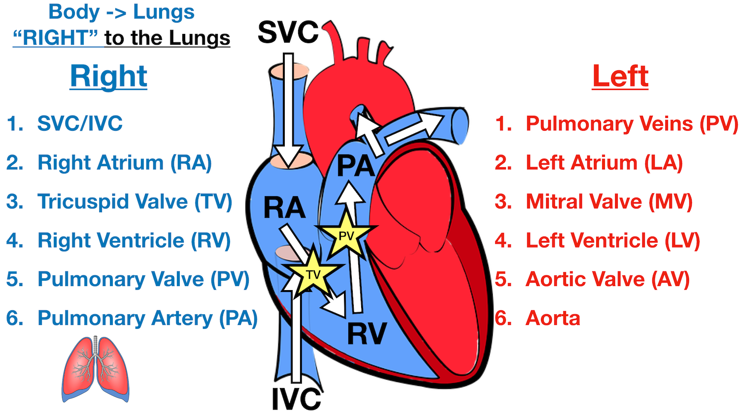
Heart Blood Flow Simple Anatomy Diagram Cardiac Circulation Pathway Steps Ezmed
At any given pressure drop the flow rate is determined by the resistance to the blood flow.

. Operation of Semilunar Valves. The body does not receive enough oxygen with these heart problems. This is a result of the left and right side of the heart working together to allow blood to flow continuously to the lungs and other parts of the body.
Capillaries of small intestine superior mesenteric vein hepatic portal vein liver sinusoids hepatic vein inferior vena cava right atrium of heart. The heart is a complex muscle that pumps blood through the three divisions of the circulatory system. The blood from the heart is carried through the body by a complex network of blood vessels Figure 1.
The job of blood is to transport oxygen and nutrients by traveling through the bodys circulatory system also called the cardiovascular system heart veins and arteries and delivering them to the other parts of the body as shown in Figure 1. Pressure drops gradually as blood flows from the major arteries through the arterioles the capillaries until blood is pushed up back into the heart via the venules the veins through the vena cava with the help of the muscles. Figure 2024 Relationships among Vessels in the Systemic Circuit.
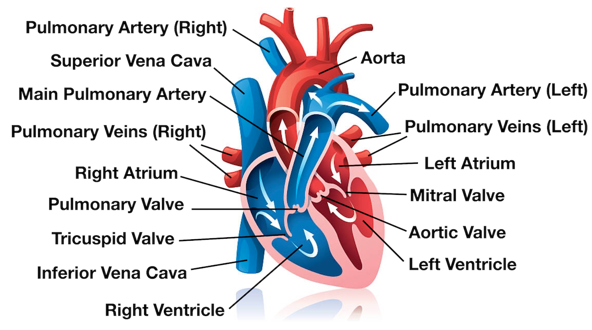
Coronary Circulation Heart is 05 of body weight but uses 5 of the circulating blood Blood vessels of heart wall nourish cardiac muscle Right left coronary arteries immediately branch off of the. Before blood flows to the various parts of the body it circulates in the heart and passes through the lungsIn this article we will describe thepath of blood through the heart. These conditions allow blood that has not been to the lungs to pick up oxygen to travel to the body.
As blood flows through the veins the rate of velocity increases as blood is returned to the heart. This condition can lead to other complications like heart disease stroke and kidney disease. Describe the structure of the heart and explain how cardiac muscle is different from other muscles.
Oxygen-poor blood enters the right side of the heart through two large veins. Explain the structure and function of venous valves in the large veins of the extremities. Trace the flow of a drop of blood from the small intestine to the right atrium of the heart noting all structures encountered or passed through on the way.
These major arteries include the carotid artery that takes blood to the brain the brachial. In the heart the two circuits of the circulatory system pulmonary and systemic circulation converge. Test your knowledge of the heart anatomy by taking up the quiz below.
The heart is charged with the sole function of pumping blood throughout the body and in so doing ensuring nutrients can be transported with ease. Normal cardiac cycles at rest take 08 seconds. It carries deoxygenated blood to the lungs where it loads up with oxygen and unloads carbon dioxide.
Problems that cause too little blood to pass through the lungs. High blood pressure is a condition in which the force of blood against the walls of blood vessels is too high. An artery is a blood vessel that carries blood away from the heart where it branches into ever-smaller vessels.
Arteries take blood away from the heart. Heart valves limit flow to a single direction. Describe the basic structure of a capillary bed from the supplying metarteriole to the venule into which it drains.
Blood flows back down the pulmonary trunk and aorta. Operation of AV Valves. Coronary circulation intrinsic to.
The heart is made of four chambers which receive and pump blood. The heart acts as a powerful pump that generates the force necessary to move the blood around the circulatory system. One heartbeat or cardiac cycle includes atrial contraction and relaxation ventricular contraction and relaxation and a short pause.
Describe the basic structure of a capillary bed from the supplying metarteriole to the venule into which it drains. The main artery is the aorta that branches into major arteries that take blood to different limbs and organs. Fills cusps to force semilunar valves closed.
Heart valve problems lead to issues with how blood flows either limiting the amount that can move. Blood is carried through the body via blood vessels. Blood is carried through the body via blood vessels.
Welcome to Anatomy of Heart Quiz. The coronary vessels that serve the heart pulmonary heart and lungs and systemic systems of the body. An artery is a blood vessel that carries blood away from the heart where it branches into ever-smaller vessels.
Babies with these forms of CHD may be cyanotic which means they look blue. The circulatory system uses the channel of blood vessels to deliver blood to all parts of the body. The relationships among blood vessels that can be compared include a vessel diameter b total cross-sectional area c average blood pressure and d velocity of blood flow.
Blood flows through the heart from veins to atria to ventricles out by arteries. In the arteries with. Explain the structure and function of venous valves in the large veins of the extremities.
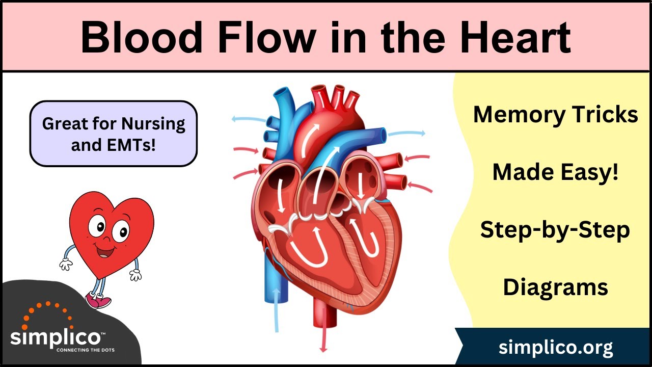
Heart Blood Flow Simple Anatomy Diagram Cardiac Circulation Pathway Steps Ezmed
The Circulatory System Before And After Birth

How Does Blood Flow Through The Heart Eett Making Movies
Heart Blood Flow Simple Anatomy Diagram Cardiac Circulation Pathway Steps Ezmed


Comments
Post a Comment